Medulloblastoma – Brief Information
Medulloblastoma is a tumour of the central nervous system (CNS). This text provides information about the characteristics and subtypes of these diseases, their frequencies, causes, symptoms, diagnosis, treatment, and prognosis.
Author: Maria Yiallouros, Editor: Maria Yiallouros, Reviewer: Dr. med. Martin Mynarek, English Translation: Dr. med. habil. Gesche Tallen, Last modification: 2024/03/29 https://kinderkrebsinfo.de/doi/e199116
Table of contents
General information on the disease
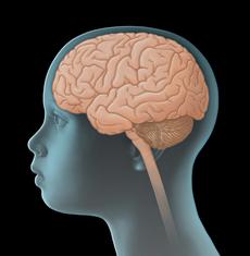
Medulloblastoma is a highly malignant, solid tumour that develops due to a malignant transformation of cells of the cerebellum, a part of the brain. Since it directly originates from the central nervous system (CNS), it is also called a primary CNS tumour, thereby differentiating it from malignant tumours of other organs that have spread (metastasised) to the CNS.
Medulloblastoma are „embryonal tumours“, which means, they originate from extremely immature (undifferentiated) cells of the central nervous system, which divide at a high rate. Therefore, these tumours grow very fast.
Generally, a medulloblastoma spreads – by uncontrolled proliferation – from the cerebellum into the adjacent tissue, for example into the brainstem, but also into the cavities of the brain (cerebral ventricles) – to be precise, into the fourth ventricle, which is located within the back part of the brain (posterior cranial fossa).
The tumour cells also spread via the cerebrospinal fluid (CSF), thereby forming metastases [see metastasis] in other parts of the central nervous system, such as the spinal canal (spinal cord). A total of one third of the patients with medulloblastoma already present with solid metastases at initial diagnosis, which are visible by imaging diagnostics. About a quarter of the patients present with tumour cells in the cerebrovascular fluid (CSF), which can be identified under the microscope. Metastasis outside the CNS, for instance to bones, bone marrow, lung, or lymph nodes, is rare for medulloblastoma.
Depending on the microscopic (histological) features as well as on molecular characteristics of the tumour, medulloblastoma are differentiated and, thus, classified into different subgroups. Their incidences and outcomes vary accordingly (see chapter „Treatment planning“).
Incidence
Approximately 3 % of all malignancies in children and adolescents are medulloblastoma. They account for about 12 % of all CNS tumours occurring in childhood and adolescence. Each year, about 60 children and adolescents under 18 years of age are newly diagnosed with medulloblastoma in Germany. This corresponds to an incidence rate of 5 per 1,000,000 children / adolescents.
Medulloblastoma are most frequent within the first nine years of life. The patients‘ average age at diagnosis is approximately seven years. Boys are affected more often than girls (gender ratio: about 2 : 1)
Causes
Medulloblastoma is caused by a malignant transformation of nerve tissue cells. The reasons for tumour development have not been completely found out yet. It is known so far, that children and adolescents with certain inherited diseases (such as Gorlin-Goltz syndrome, Li-Fraumeni syndrome, or Fanconi anaemia) have a higher risk of developing a medulloblastoma than their healthy peers. Since these genetic conditions are associated with an elevated risk for tumour development, they are also known as cancer predisposition syndromes.
In addition, it has been shown that medulloblastoma are frequently associated with certain chromosomal aberrations within cells. The resulting impairments of cell development and cell communication may be contributing factors promoting the transformation of a healthy into a cancer cell. Also, radiation therapy of the brain, for example as received by patients with certain forms of leukaemia or with eye cancer (retinoblastoma), may lead to an increased risk of developing a CNS tumour later in life.
Symptoms
Due to the uncontrolled and aggressive growth pattern of medulloblastoma, symptoms typically develop and deteriorate fast. Similar to those of other tumours of the central nervous system (CNS), the presenting symptoms of medulloblastoma primarily depend on the patient’s age, the site and size of the tumour as well as its pattern of spread within the CNS. The following general (nonspecific) and local (specific) symptoms can occur:
General (nonspecific) symptoms
Unspecific general symptoms occur independently of the tumour’s location. They may be similar to and therefore mimic other, non-CNS diseases. General symptoms of a child or adolescent with a CNS tumour may include headaches and/or back pain, dizziness, loss of appetite, nausea and vomiting (particularly after getting up in the morning), weight loss, increasing fatigue, inability to concentrate, school problems, mood swings, and character changes as well as developmental delay, to name a few.
Major reason for these symptoms is the slowly but continuously increasing intracranial pressure (ICP). An elevated intracranial pressure may be caused by the growing, thus more and more space-occupying tumour within the bony skull. It may as well be due to the tumour blocking the regular flow of the cerebrospinal fluid – as is frequent in medulloblastoma patients –, thereby forming hydrocephalus. In babies or small children with soft spots (open fontanelles), elevated intracranial pressure and hydrocephalus typically present with a bulging fontanelle or a larger than expected head circumference (macrocephalus), respectively.
Local (specific) symptoms
Local symptoms may indicate the tumour location and, thus, which functional regions of the CNS might be affected. Thus, tumours like medulloblastoma, which arise in the cerebellum and frequently spread into the into the fourth ventricle and the brainstem, can cause dizziness and gait disturbances, increasing imbalance including insecurity when jumping or walking stairs as well as sensory and coordination problems.
Also, visual deficits (such as strabism, double vision, and uncontrolled eye movements) due to impaired cranial nerves can occur. In case medulloblastoma has spread to other areas of the central nervous system, other symptoms may occur, such as back pain or muscle weakness when the spinal cord is affected.
Good to know: Not all patients presenting with one or more of the symptoms mentioned above do have a medulloblastoma or another type of brain tumour. Many of these symptoms may also occur with other, harmless diseases that are not associated with a brain tumour at all. However, if certain symptoms persist or get worse (for example repetitive headaches or rapid increase of head circumference in a young child), a doctor should be seen to find the underlying reason. In case it is a medulloblastoma or some other brain tumour, treatment should be started as soon as possible.
Diagnosis
If the paediatrician thinks that the young patient’s history (anamnesis) and physical examination are suspicious of a tumour of the central nervous system (CNS), the child should immediately be referred to a hospital with a childhood cancer program (paediatric oncology unit), where further diagnostics can be initiated and performed by childhood cancer professionals. Very close collaboration between various specialists (such as paediatric oncologists, paediatric neurosurgeons, paediatric radiologists, to name a few) is required, both to find out, whether the patient really suffers from a malignant CNS tumour and, if so, to determine the tumour type and the extension of the disease. Knowing these details is absolutely essential for optimal treatment and prognosis.
Tests to secure diagnosis
The initial diagnostic procedures for a young patient presenting with a suspected CNS tumour at a childhood cancer centre include another assessment of the patient’s history, a thorough physical/neurological exam and imaging diagnostics, such as magnetic resonance imaging (MRI) or (less often) computed tomography (CT). These tests help find out exactly whether the patient has a tumour of the central nervous system. Also, localisation and extent of the tumor, its demarcation regarding adjacent tissue as well as a potential hydrocephalus can be diagnosed by these imaging techniques very well.
In order to validate the final diagnosis, histological and molecular analysis of surgically obtained tumour tissue (biopsy) is required. Usually, this is done using the tissue obtained during surgical tumour removal.
The extent of histological and, especially, molecular genetic workup has been substantially increased over the past years. Today’s option of using modern lab techniques makes it possible to identify molecular tissue characteristics that do not only help finalize the diagnosis, but can also provide information on what to expect regarding the course of the disease (such as growth behaviour). Hence, molecular diagnostics already play an important role in treatment planning and will most certainly become even more relevant in the future.
Tests to assess spread of disease
Once the diagnosis of a medulloblastoma has been confirmed, additional tests are required to assess the extent of the disease within the central nervous system (CNS). Apart from MRI scans of the complete CNS (brain and spine) for macroscopic metastases [see metastasis], these tests also include microscopic checking of the cerebrospinal fluid (CSF) for tumour cells in the spinal cord (which are not visible by MRI scan). Cerebrospinal fluid is mostly obtained from the spine in the lower back (lumbar puncture), since the risk of the puncture needle damaging the spinal cord is lowest at the lower back level.
Tests before treatment begins
In preparation for the intensive treatment of the brain tumour, further investigations are performed, such as electrocardiography (ECG) and echocardiography to check cardio function. Furthermore, additional blood tests are needed to assess the patient’s general health condition and to check whether the function of certain organs (such as liver and kidneys) is affected by the disease and whether there are any metabolic disorders to be considered prior or during therapy. Any changes occurring during the course of treatment can be assessed and managed better based on the results of those initial tests, which thus help to keep the risk of certain treatment-related side effects as low as possible.
Also, the patient’s blood group needs to be determined in case a blood transfusion is required during treatment. In sexually mature females (which means after they have experienced their first menstrual bleeding), a pregnancy test is recommended prior to treatment as well.
Good to know: Not every patient needs the complete check-up. On the other hand, tests might be added that haven't been mentioned here, depending on the individual situation of the patient. Your caregivers will inform you and your child, which diagnostic procedures are individually required in your case and why.
Treatment planning
Once the diagnosis and extent of a medulloblastoma has been confirmed, treatment planning starts. In order to provide the patient with the best possible individual and risk-adapted therapy, the treatment team considers specific factors that are known to have an impact on the prognosis (so-called prognostic factors). One important prognostic factor is the type (subtype) of medulloblastoma, since each subtype correlates with a different growth pattern and malignity. Further prognostic factors are the size, localization, and spread of the tumour (metastasis), for these factors have an impact on whether or not the tumour can be completely removed by means of an operation, thus influencing the patient’s chances of survival.
In addition, response to chemotherapy and the patient’s age at the time point of diagnosis play an important role. Age at diagnosis, for example, determines whether the patient may receive radiotherapy or not. Also, cancer predisposition syndromes (such as Li-Fraumeni syndrome, Gorlin-Goltz syndrome, or Fanconi anaemia) and the patient’s overall physical condition are of importance.
All these factors are included in treatment planning in order to achieve the best outcome possible for each patient.
Classification of Medulloblastoma
According to the World Health Organization (WHO), medulloblastoma is defined as a high-grade malignant tumour (WHO grade IV). However, there are various subtypes of medulloblastoma: They look different under the microscope, meaning histologically, and also present with different molecular characteristics. Since these differences are sometimes associated with different prognostic outcomes, too, considering them is crucial for optimal treatment planning.
As per classification of the World Health Organization (WHO classification) for tumours of the central nervous system, the following subtypes of medulloblastoma have been defined:
- classic Medulloblastoma (CMB)
- desmoplastic/nodular Medulloblastoma (DMB)
- Medulloblastoma with extensive nodularity (MBEN)
- anaplastic Medulloblastoma (AMB)
- large cell Medulloblastoma (LC MB)
However, the appearance of the tumours under the microscope has turned out not to be insufficient to predict their growth behaviour. Therefore, since 2016 classification has been based on molecular genetic (biological) tumour characteristics in addition to the histological features. This additional consideration of molecular factors has shown to provide a more precise assessment of the tumour biology and, thus, is more feasible for optimal treatment planning. The WHO currently classifies four genetic (molecular) Medulloblastoma subgroups: WNT-activated, SHH-activated, group 3 and group 4.
Histological and/or molecular medulloblastoma subgroups with either favourable or unfavourable prognosis exist. For example, patients with desmoplastic/nodular medulloblastomas and medulloblastomas with extensive nodularity usually have a more favourable outcome than patients with other histological subtypes. Same for the WNT-activated medulloblastoma, which can be associated with a better prognosis when compared to a group 3 medulloblastoma, especially when the latter harbours a genetically unfavourable alteration (MYCC or MYCN amplification).
Every patient will be stratified – based on all prognostic factors (histological/molecular subtype, metastasis, residual tumour, age at diagnosis) – into a specific treatment group (such as low-risk, standard risk or high-risk group), each of which considers the individual risk of relapse. The higher the risk of relapse, the more intense is usually the treatment.
Treatment
Treatment of children and adolescents with medulloblastoma should take place in a children's hospital with a paediatric oncology program. Only in such a childhood cancer centre, highly experienced and qualified staff (doctors, nurses and many more) is guaranteed, since they are specialized and focus on the diagnostics and treatment of children and teenagers with cancer according to the most advanced treatment concepts. The doctors in these centres collaborate closely with each other. Together, they treat their patients according to treatment plans (protocols) that are continuously optimised. The goal of the treatment is to achieve higher cure rates while avoiding side effects as much as possible.
Current treatment concepts involve neurosurgical tumour removal, chemotherapy and, depending on the patient’s age, radiotherapy of the brain and spinal cord. For some patients, high-dose chemotherapy followed by stem cell transplantation may be an option, too.
Surgery
Immediate surgery for tumour removal is crucial for patients with medulloblastoma, because most of them are in critical clinical condition due to the tumour and subsequent impairment of cerebrovascular fluid flow (which can cause hydrocephalus). Goal of surgery is gross (microsurgical) total tumour resection. This means that at the end of the surgical procedure, no tumour tissue can be identified through the surgical microscope.
Due to these microsurgical techniques [neurosurgery], total tumour resection can be achieved for more than 50 % of patients with medulloblastoma today. If – at the beginning of treatment – the tumour cannot be completely removed without risking a substantial damage of the healthy brain tissue, a second attempt of complete resection may be evaluated at a later time point, for example after radio- and/or chemotherapy.
In most medulloblastoma patients, tumour removal also results in normalising the flow of cerebrospinal fluid (CSF). Patients initially presenting with hydrocephalus may need a transient hydrocephalus drainage prior to tumour removal or, sometimes, even a permanent drainage system later.
Additional, non-surgical treatment
Since medulloblastomas tend to infiltrate adjacent tissue and, furthermore, often spread into other parts of the central nervous system via the cerebrovascular fluid (CSF), only treating the tumour locally is not sufficient. Therefore, surgery is followed by additional non-surgical treatment, comprising chemotherapy and, partly, radiation therapy.
Chemotherapy uses drugs (so-called cytostatic agents or cytostatics) that can kill fast-dividing cells, such as cancer cells, or inhibit their growth, respectively. Radiotherapy is done using energy-rich, electromagnetic radiation, given through the skin to the tumour region. Radiation causes DNA damage in tumour cells, thereby leading to cell death. Aside from this so-called conventional radiotherapy, particle-radiation with protons (also known as proton therapy) can be an option for some patients as well. This type of radiotherapy provides the benefits of better targeting the tumour area, thus sparing more adjacent, healthy tissue from the effects of radiation. Proton therapy is gaining an increasing importance in the treatment of children and teenagers with solid tumours.
Decision upon which therapy is to be applied (treatment modalities and intensity) is based on the patient’s age, the histological and molecular subtype of the tumour, certain biological risk factors as well as on the extent of both metastases and surgical tumour removal.
The following paragraphs provide an overview of various treatment options. Please note, that – due to the complexity of treatment options (according to the large spectrum of molecular subtypes and other relevant risk factors) – we do not claim completeness. Your child’s caregiver team will provide you with the details applying to the best treatment options for your child. In general, high-risk patients receive a more intense therapy than patients with low- or standard-risk medulloblastoma.
Treatment of low- and standard-risk Medulloblastoma in patients older that 3-5 years
Children and adolescents with non-metastasised medulloblastoma, who – based on the tumour biology – have been stratified into the low- or standard-risk group, respectively, and who are older than 3-5 years at diagnosis (the age limit is adjusted to the medulloblastoma subtype), usually receive radiotherapy of the complete central nervous system (craniospinal radiotherapy), followed by an additional radiation boost to the tumour site. Radiotherapy is followed by a so-called maintenance chemotherapy, which includes a combination of cytostatic agents. Medulloblastoma patients of both risk groups can be enrolled in the therapy optimising trials SIOP-PNET 5 MB (LR/SR-arm) (see chapter "Therapy optimising trials and registries").
Treatment of high-risk Medulloblastoma in patients older that 3-5 years
Patients over 3-5 years with metastasised disease and/or who have been stratified to the high-risk group based on the biology of their tumour, will usually receive an intensified treatment. Depending on the individual risk factors, an intensified regimen may include radiotherapy with higher doses and, for some patients, even a so-called induction chemotherapy, which is given prior to radiotherapy. Either or, radiotherapy (of central nervous systems and tumour site) is always followed by maintenance chemotherapy. Since 2018, certain high-risk patients are eligible to participate in the therapy optimising trial SIOP-PNET 5 MB) (see chapter Therapy optimising trials and registries).
Treatment of children younger than 3-5 years
Brain development is ongoing in children younger than 3 to 5 years of age. Therefore, in order to prevent or at least minimise the risk of severe late effects, current treatments do not routinely include radiotherapy for this age group or consider it as a delayed treatment option. Instead, surgical tumour removal is followed by chemotherapy with multiple agents. Chemotherapy can differ in intensity, depending on the tumour type and individual risk status (standard-/high-risk medulloblastoma).
Subsequent therapy, as well, is based on the tumour type, existing metastasis and patient’s age at diagnosis. Standard-risk patients with good response to treatment (that means complete or partial tumour elimination) usually continue with chemotherapy. In case of no or insufficient tumour shrinkage, children older than 18 months of age may receive radiotherapy of the tumour region. In younger children, time until they are 18 months old is bridged by another round of chemotherapy. Also, a second surgery to remove residual tumour is regularly being considered.
Some patients may also benefit from high-dose chemotherapy followed by autologous stem cell transplantation to increase their chances of survival. Current treatment regimens particularly consider this option for children with metastasised medulloblastoma who are younger than four years, as well as for certain patients with recurrent disease (recurrence, relapse).
Treatment in case of relapse
The treatment regimens for patients with relapse are generally considering the patient’s overall condition, the intensity of prior treatment, and the initial response to chemotherapy. In general, the treatment of recurrent disease includes all options of local treatment (surgery, radiotherapy) as well as chemotherapy.
Therapy optimising trials and registries
The majority of the children and adolescents with medulloblastoma or a relapse of this disease receive therapy according to the treatment plans of therapy optimising trials or registries.
Therapy optimising trials are standardised and controlled clinical trials that aim at steadily developing and improving treatment concepts for sick patients based on the current scientific knowledge. Patients who cannot participate in any study, for example because none is available or open for them at that time, or since they do not meet the required inclusion criteria, respectively, are often included in a so-called registry. One goal of a registry is to collect treatment data for research questions. Furthermore, the registry center supports the doctors at site with (non-commital) treatment recommendations based on the most recent data on best treatment options, in order to provide the patient with optimal therapy even without the framework of a clinical study.
Currently, the following therapy optimising trials / registries are available for patients with medulloblastama in Germany:
- Trial SIOP-PNET 5 MB for patients with medulloblastoma: Since April 2014, patients with medulloblastoma can participate in the European-wide conducted study SIOP-PNET 5 MB. Initially open only for patients with low- or standard-risk and recurrent disease, the study has been offering different treatment arms also for certain high-risk patients since fall 2018. Aside from patients with WNT-activated medulloblastoma, who – due to age, metastasised disease and/or residual tumour – harbour a high risk of recurrent disease (WNT-HR), these also include patients with certain hereditary cancer predisposition syndromes (SHH medulloblastoma with TP53-mutation). SIOP-PNET 5 MB also involves an open registry for patients who are not eligible to be recruited to one of the study arms, such as patients with other types of cancer predisposition syndromes. One of the eligibility criteria for both study and registry is an age of 3-5 years at diag-nosis (depending on the medulloblastoma subtype). The headquarters of the trial are located in the Children’s Cancer Centre at the University of Hamburg, Germany. The head of the trial is Prof. Dr. med. Stefan Rutkowski.
- I-HIT-MED Registry: Patients with medulloblastoma, who, for different reasons, cannot or do not want to participate in any currently available or open trial, can be en-rolled in this registry (International HIT-MED Registry), regardless of the treatment given. These patients will receive treatment as per individually designed treatment plans. The goal of the registry is not to assess the feasibility of an ongoing trial, safety or efficacy of a certain treatment. It rather aims at collecting individual patient data for future analysis. The headquarters of the registry are located in the Children’s Cancer Centre at the University of Hamburg, Germany (head of study: Prof. Dr. med. Stefan Rutkowski).
- HIT-REZ Registry: Patients, whose disease does not respond to current treatments (therapy-resistant, progressive medulloblastoma) or with recurrent disease (relapse), respectively, can be enrolled in this registry, which has been open since January 2015. This registry does not serve to test new treatment regimens or drugs. However, the experts running the registry are providing treatment recommendations based on the most recent results obtained from national trials (for example from the HIT-REZ 2005 trial, which was closed in 2016) as well as international relapse trials. The headquarters of the registry are located in the Children’s Cancer Centre at the University of Essen, Germany. The head of the study is Prof. Dr. med. Gudrun Fleischhack.
Prognosis
The chances of cure (prognosis) for children and adolescents with medulloblastoma have improved considerably over the last decades. Today’s modern diagnostic procedures and the use of intensive, standardised combination therapies currently result in 5-year survival rates of about 75 % and 10-year survival rates of almost 70 %.
Individual prognosis is influenced by the medulloblastoma subtype, existence of metastasis, age at diagnosis and response to therapy. Patients with a favourable risk profile can achieve survival rates higher than 80%, particularly patients with desmoplastic/nodular and WNT-activated medulloblastoma. Patients with unfavourable risk profile (such as with anaplastic or large-cell medulloblastoma or with MYCC or MYCN amplification) have a lower chance of cure. Metastases can (though not necessarily always) be associated with a more unfavourable outcome, since prognostic co-factors such as tumour type and treatment response have an impact, too.
Until the end of the Eighties, prognosis of young children (younger than 4 years) was significantly poorer than in older children and adolescents. It improved substantially after the introduction of treatment protocols with intensified chemotherapy. Patients with recurrent disease (recurrence) usually have a rather unfavourable prognosis. In individual settings, chances of long-term survival can be achieved by high-dose chemotherapy followed by autologous stem cell transplantation.
Note: The survival rates mentioned in the text above are statistical values. Therefore, they only provide information on the total cohort of patients with childhood medulloblastoma. They do not predict individual outcomes.
In the context of cancer, the term „cure“ should rather be referred to as „free of cancer“, for even if current treatment regimens may help remove the tumour, the the tumour’s growth may have caused irrepairable damage to the brain or the treatment may be associated with late-effects. Early detection and appropriate management of these long-term secondary effects typically require intensive rehabilitation and thorough long-term follow-up care, although a patient may have been „cured“ from the cancer.
 PDF Information on Medulloblastoma (274KB)
Literatur 
- Erdmann F, Kaatsch P, Grabow D, Spix C: German Childhood Cancer Registry - Annual Report 2019 (1980-2018). Institute of Medical Biostatistics, Epidemiology and Informatics (IMBEI) at the University Medical Center of the Johannes Gutenberg University Mainz 2020 [URI: https://www.kinderkrebsregister.de/ typo3temp/ secure_downloads/ 42507/ 0/ 1c5976c2ab8af5b6b388149df7182582a4cd6a39/ Buch_DKKR_Jahresbericht_2019_komplett.pdf]
- Fleischhack G, Rutkowski S, Pfister SM, Pietsch T, Tippelt S, Warmuth-Metz M, Bison B, van Velthoven-Wurster V, Messing-Jünger M, Kortmann RD, Timmermann B, Slavc I, Witt O, Gnekow A, Hernáiz Driever P, Kramm C, Benesch M, Frühwald MC, Hasselblatt M, Müller HL, Sörensen N, Kordes U, Calaminus G: ZNS-Tumoren. in: Niemeyer C, Eggert A (Hrsg.): Pädiatrische Hämatologie und Onkologie. Springer-Verlag GmbH Deutschland, 2. vollständig überarbeitete Auflage 2018, 359 [ISBN: 978-3-662-43685-1]
- Rutkowski S: Medulloblastom im Kindes- und Jugendalter. Leitlinie der Gesellschaft für Pädiatrische Onkologie und Hämatologie. AWMF online 2018 [URI: http://www.awmf.org/ uploads/ tx_szleitlinien/ 025-009l_S1_Medulloblastom-Kinder-Jugendliche_2018-04.pdf]
- Ripperger T, Bielack SS, Borkhardt A, Brecht IB, Burkhardt B, Calaminus G, Debatin KM, Deubzer H, Dirksen U, Eckert C, Eggert A, Erlacher M, Fleischhack G, Frühwald MC, Gnekow A, Goehring G, Graf N, Hanenberg H, Hauer J, Hero B, Hettmer S, von Hoff K, Horstmann M, Hoyer J, Illig T, Kaatsch P, Kappler R, Kerl K, Klingebiel T, Kontny U, Kordes U, Körholz D, Koscielniak E, Kramm CM, Kuhlen M, Kulozik AE, Lamottke B, Leuschner I, Lohmann DR, Meinhardt A, Metzler M, Meyer LH, Moser O, Nathrath M, Niemeyer CM, Nustede R, Pajtler KW, Paret C, Rasche M, Reinhardt D, Rieß O, Russo A, Rutkowski S, Schlegelberger B, Schneider D, Schneppenheim R, Schrappe M, Schroeder C, von Schweinitz D, Simon T, Sparber-Sauer M, Spix C, Stanulla M, Steinemann D, Strahm B, Temming P, Thomay K, von Bueren AO, Vorwerk P, Witt O, Wlodarski M, Wössmann W, Zenker M, Zimmermann S, Pfister SM, Kratz CP: Childhood cancer predisposition syndromes-A concise review and recommendations by the Cancer Predisposition Working Group of the Society for Pediatric Oncology and Hematology. American journal of medical genetics. Part A 2017, 173: 1017 [PMID: 28168833]
- Louis DN, Perry A, Reifenberger G, von Deimling A, Figarella-Branger D, Cavenee WK, Ohgaki H, Wiestler OD, Kleihues P, Ellison DW: The 2016 World Health Organization Classification of Tumors of the Central Nervous System: a summary. Acta neuropathologica 2016, 131: 803 [PMID: 27157931]
- Sabel M, Fleischhack G, Tippelt S, Gustafsson G, Doz F, Kortmann R, Massimino M, Navajas A, von Hoff K, Rutkowski S, Warmuth-Metz M, Clifford SC, Pietsch T, Pizer B, Lannering B, SIOP-E Brain Tumour Group: Relapse patterns and outcome after relapse in standard risk medulloblastoma: a report from the HIT-SIOP-PNET4 study. Journal of neuro-oncology 2016, [PMID: 27423645]
- Rutkowski S, Trollmann R, Korinthenberg R, Warmuth-Metz M, Weckesser M, Krauss J, Pietsch T: Leitsymptome und Diagnostik der ZNS-Tumoren im Kindes- und Jugendalter. Gemeinsame Leitlinie der Gesellschaft für Neuropädiatrie und der Gesellschaft für Pädiatrische Onkologie und Hämatologie 2016 [URI: https://register.awmf.org/ assets/ guidelines/ 025-022l_S1_ZNS-Tumoren_Kinder_Jugendliche_2016-09-abgelaufen.pdf]
- Gerber NU, Mynarek M, von Hoff K, Friedrich C, Resch A, Rutkowski S: Recent developments and current concepts in medulloblastoma. Cancer treatment reviews 2014, 40: 356 [PMID: 24389035]
- Lannering B, Rutkowski S, Doz F, Pizer B, Gustafsson G, Navajas A, Massimino M, Reddingius R, Benesch M, Carrie C, Taylor R, Gandola L, Björk-Eriksson T, Giralt J, Oldenburger F, Pietsch T, Figarella-Branger D, Robson K, Forni M, Clifford SC, Warmuth-Metz M, von Hoff K, Faldum A, Mosseri V, Kortmann R: Hyperfractionated versus conventional radiotherapy followed by chemotherapy in standard-risk medulloblastoma: results from the randomized multicenter HIT-SIOP PNET 4 trial. Journal of clinical oncology 2012 Sep 10; 30: 3187 [PMID: 22851561]
- Frühwald MC, Rutkowski S: ZNS-Tumoren bei Kindern und Jugendlichen. Dtsch Arztebl Int 2011; 108: 390
- Rutkowski S, Cohen B, Finlay J, Luksch R, Ridola V, Valteau-Couanet D, Hara J, Garre ML, Grill J: Medulloblastoma in young children. Pediatr Blood Cancer 2010, 54: 635 [PMID: 20146217]
- von Hoff K, Hinkes B, Gerber NU, Deinlein F, Mittler U, Urban C, Benesch M, Warmuth-Metz M, Soerensen N, Zwiener I, Goette H, Schlegel PG, Pietsch T, Kortmann RD, Kuehl J, Rutkowski S: Long-term outcome and clinical prognostic factors in children with medulloblastoma treated in the prospective randomised multicentre trial HIT'91. European journal of cancer (Oxford, England : 1990) 2009, 45: 1209 [PMID: 19250820]
- Rutkowski S, Bode U, Deinlein F, Ottensmeier H, Warmuth-Metz M, Soerensen N, Graf N, Emser A, Pietsch T, Wolff JE, Kortmann RD, Kuehl J: Treatment of early childhood medulloblastoma by postoperative chemotherapy alone. The New England journal of medicine 2005, 352: 978 [PMID: 15758008]