Structure and function of the kidneys
Author: Maria Yiallouros, Editor: Maria Yiallouros, English Translation: Dr. med. habil. Gesche Tallen, Last modification: 2022/02/04 https://kinderkrebsinfo.de/doi/e128812
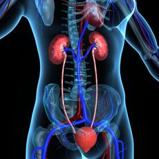
A healthy human being has two kidneys. These are bean-shaped organs located in the rear abdominal cavity, left and right of the spine, slightly beneath the diaphragm under the rib cage, with the left kidney sitting behind the spleen and the right one behind the liver.
The concave border of each kidney has a recessed area that is called the renal (renal means "kidney-associated") hilum. This is where the renal artery enters to supply the kidney with blood carrying oxygene and nutrients, but also waste products, and where the renal vein leaves, carrying the blood that was filtered by the kidney. The hilum is also the entry for lymph vessels, nerves and the ureter. The ureter is a muscular tube that transports the urine from the kidney to the urinary bladder, from where it is released from the body.
Each kidney is encapsulated by a solid cover of connective tissue (renal capsule) and embedded in further layers of fat and connective tissue, which protect the organ from trauma and anchor it to the rear abdominal wall
The inner organ can be divided into three major parts: the outer renal cortex, the inner renal medulla and the renal pelvis in the area of the hilum.
Good to know: major function of the kidneys is to filter the blood from toxins and metabolic end products as well as to remove and retain water and electrolytes by making urine, which is released via the ureter and the urinary bladder.
(© 7activestudio - Fotolia.com)
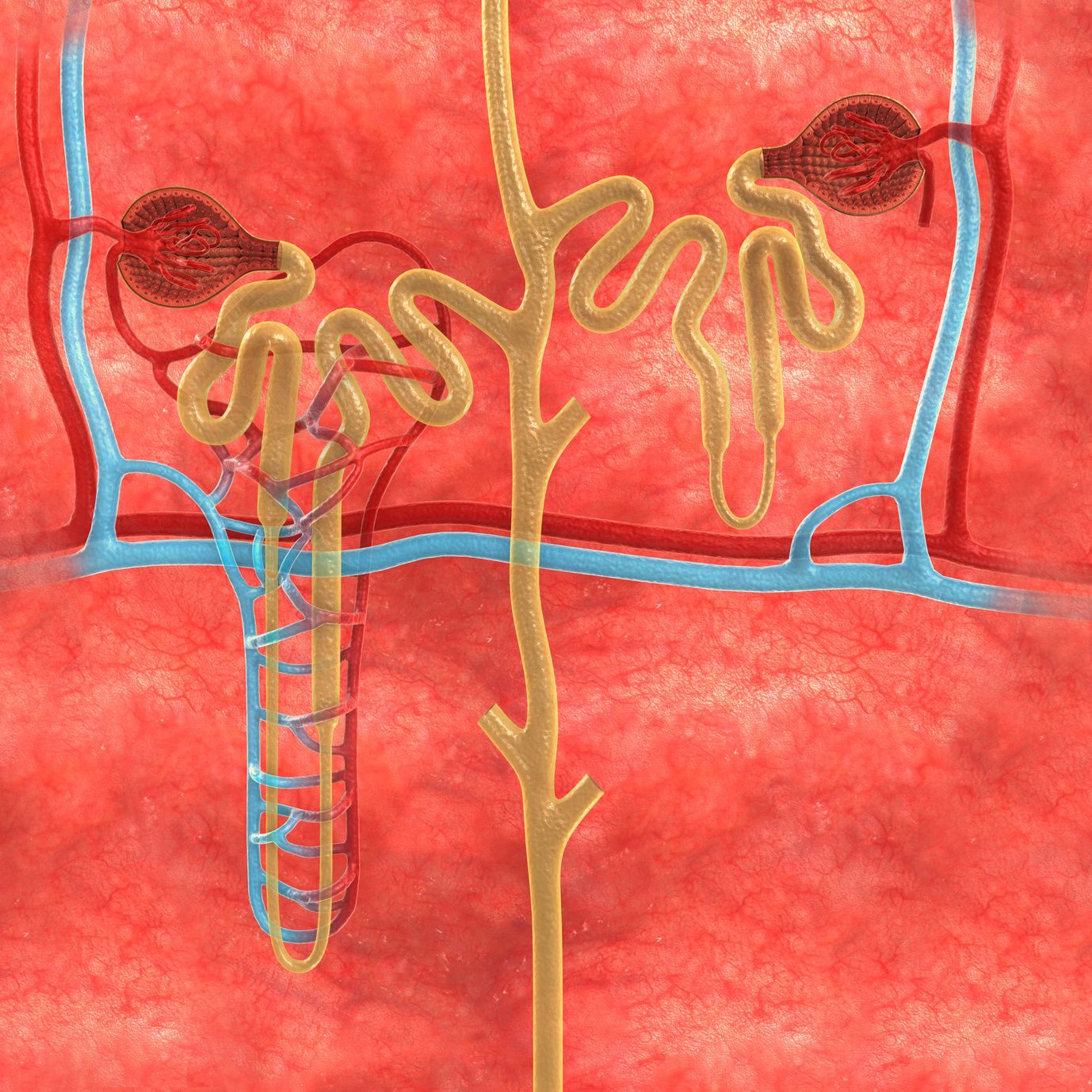
Kidney tissue with nephrons
The kidney tissue (parenchyma) contains about one million little filter units, the so-called nephrons. These nephrons filter the blood and produce the urine. Each nephron consists of a so-called renal corpuscule followed by a tubule.
Together, corpuscule and tubule form a functional unit: the corpuscules filter the blood, thereby producing about 100 litres of urine every day. To reduce this large volume, the tubules regain most of the filtered electrolytes and fluids (about 99 %) and return it to the blood circulation. As a result, only about a tenth of the initially produced urine (called primary urine), for example 1 - 1.5 liters, is finally released.
(The volume of the primary and final urine produced by the corpuscules per day mainly depends on the fluid intake as well as on the total blood volume, which is higher in adults than in children.)
While the renal corpuscules are found in the outer renal cortex, the tubules are located within the inner renal medulla and drain – via special collecting ducts – into the so-called renal calyces. This is where the urine is collected and transported to the renal pelvis, ureter and urinary bladder.
Overview of the kidneys' chores
- draining the blood from metabolic waste – such as as uric acid, urea (nitrogen) and creatinine – and from toxins (for example medication) by making urine that is released via the urinary tract
- controlling the body's fluid balance, thereby regulating blood pressure
- controlling mineral and acid-base balance (by regulating the urine's composition)
- secreting hormones, such as erythropoietin for stimulating the production of blood cells, and renin for regulating blood pressure
- excretion of peptide hormones